A UW-Madison professor developed a way to more easily distinguish between different substances. Now, she’s been given a $480,135 grant by the National Eye Institute to study how her technique can be used in the detection of eye diseases.

Have you ever started with an idea that at first seemed inconsequential, but over time it grew into something extraordinary? That was the case for Dr. Melissa Skala, an investigator at the Morgridge Institute for Research and a professor of biomedical engineering at UW-Madison. It all started with an idea for a method that makes use of the photothermal effect to create contrast — that is, to make it easy to distinguish between one substance and another. The idea is that if you point a very low-power laser at a substance, it will cause the substance to produce a small amount of heat. Then, an interferometer (a device that’s very good at measuring temperature change) can detect the heat, allowing a scientist to focus on the substance itself and ignore the background. “We turn it on and off really quickly, and that’s how we become sensitive to where the absorption is,” Skala says. “That helps us reject the background.”
Though proud of her technique, Skala admits that at first, she didn’t think there were any practical uses for it. “I actually started this technology development as a postdoc, and it was just this one-off, crazy idea… so people were like, ‘Oh, that’s cute,’” Skala recalls. But when she crossed paths with Dr. Joseph Carroll, a professor of ophthalmology (the branch of medicine that deals with the diagnosis and treatment of eye diseases) and the Director of the Advanced Ocular Imaging Program at the Medical College of Wisconsin, he immediately saw the usefulness of her method. “[Joe] really convinced me and pushed this work forward, because he’s a person who works on eye disease, and he needs tools that will help him understand and treat it better,” Skala recalls. “It almost takes someone who sees the potential of what you’re doing to show you it can be useful, and that’s what happened here.”
What Carroll saw in Skala’s technique was its potential to be used to take pictures of the different layers of the retina. What’s different about Skala’s method is that it also allows the ophthalmologist to consider the different molecular features of the eye. “The problem [with existing techniques] is they’re like ultrasound in that they give you structural information… but they don’t provide any molecular information, so you just get the skeleton, basically. And so, this technique allows us to understand how the eye functionally changes, not just structurally changes,” Skala says. “It adds a functional component to something that already exists.”
This molecular information is especially useful in that it can provide insight into how substances such as the pigment melanin accumulate in the eye. Change in melanin distribution is an early warning sign for diseases such as age-related macular degeneration, a common condition among older adults that leads to vision loss. “Currently, we can only detect that disease once you’re starting to lose your vision, and the best we can do is try to stop vision loss,” Skala says. “But if we can detect it before then, hopefully we can avoid any vision loss.”
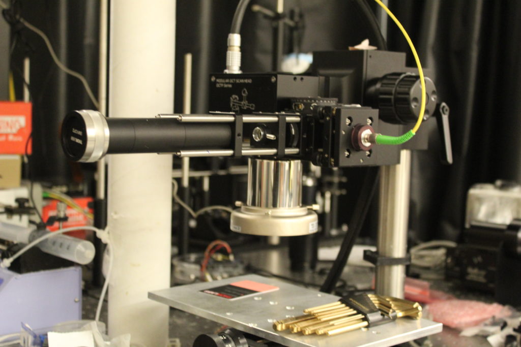
Photo by: James Ballard
Skala also sees the technique having useful applications in detecting cancer and in aiding certain surgical procedures. She hopes that once human studies for the technique conclude (the project has already been approved by the FDA and is now being supported by a $480,135 grant from the National Eye Institute), it will be widely applied to detecting all sorts of eye diseases. “If it works out, I think it could be extremely helpful for many eye diseases. It’s not what we developed it for, but I think the technology finds its usefulness in its own way,” Skala says.
Nevertheless, Skala anticipates that there will be challenges along the way. “We still have to optimize the speed that we can collect the images, which is challenging,” Skala concedes. Her team will also need to ensure that their technology can distinguish between melanin and other materials such as lipofuscin, a brown-yellow pigment that also accumulates when eye diseases are present. However, she remains optimistic that her team will find a way to overcome these hurdles. “I guess that’s the point of the grant. The NIH took a risk — this is a high-risk, high-reward kind of grant,” Skala says. If her previous technological triumphs are any indicator, she will meet these challenges and “see” them all the way through.